Poster Presentation The Annual Scientific Meeting of the Endocrine Society of Australia and the Society for Reproductive Biology 2014
Do sheep have pelvic organ prolapse and differences in vaginal pressure? (#230)
Sheep develop spontaneous POP at a similar frequency as women and maybe a suitable model for preclinical studies evaluating cell-based therapies for POP. The aim was to establish a clinical score of POP in ewes and correlate this with vaginal tone measured with a fibre-optic pressure sensor device.
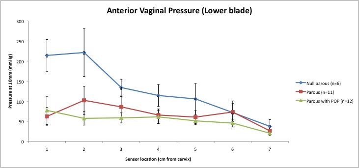
Vaginal examinations were conducted on 29 conscious ewes (6 nulliparous, 23 parous) without sedation standing in a V drive, by adapting the human POP-Q (1). Maximum displacement of vaginal tissue at POP-Q points; Aa, 3cm above the introitus on the anterior vaginal wall (range -3above to +3 below the introitus); Ap, 3cm above the introitus (posterior wall); Ba above the urethra (anterior wall) (-3 to total vaginal length);GH (genital hiatus); PB (perineal body). Ovine POP was defined as descent to the introitus, or increased Ba (0) relative to the urethra. The pressure sensor device has 7 pressure sensors on each blade of a speculum, which electronically recorded vaginal pressures at 10mm dilation.
There was no evidence of tissue mobility at Aa, Ap, Ba in nulliparous ewes, which also had smaller GH and PB compared to parous ewes (Table 1). Twelve parous sheep showed significant POP at Ba, with greater tissue mobility at Aa and Ap compared to the other parous ewes (P< 0.0001, 0.003, and 0.019 respectively). Nulliparous ewes showed significantly greater pressures in the upper vagina (Ba, sensors 1,2,3) (P<0.0001) compared to parous sheep with/without POP. Parous ewes with POP had lower pressures at point 3 compared to those without (Figure 1&2).
Our data suggests correlation between increased vaginal tissue mobility and lower vaginal tone in the upper anterior vagina of parous ewes that is not evident in nulliparous animals. These methods may predict subclinical POP in sheep enabling their selection for assessing new treatments for POP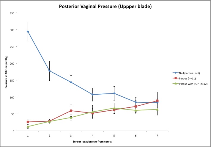

- 1. Bump RC, Mattiasson A, Kari B, Brubaker LP, Delancey JOL, Klarskov P, et al. The standardization of terminology of female pelvic organ prolapse and pelvic floor dysfunction. 1996;10–7.